
So, the neurons are the structural and trophic unit of the nervous system that transform and sustain what they innervate. Structure of neuron under light microscope

And the supportive cells of the peripheral nervous system (PNS) include Schwann and Satellite cells. And you already know that the neuron is the structural and functional unit of the nervous system.Īgain, the supportive or glial cells of the nervous tissue consist of neuroglial and ependymal cells in the central nervous system (CNS). Observing the nervous tissue under the light microscope will find neurons (nerve cells) and supportive (glial cells) in its parenchyma. I hope you can understand what nervous tissue is.

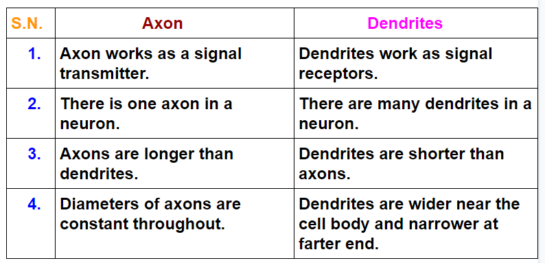
Presence of a relatively long, cylindrical axon process that arises from the neuron’s axon Hillock (shown in slide image diagram).There are highly branched short dendritic processes present around the neuron’s cell body.Presence of easily visible cytoplasm and plasma membrane (neurilemma) in the body of a neuron.The cell body possesses spherical, euchromatic, and large eccentric nuclei containing a prominent nucleolus.Presence of an identifiable cell body (soma) that locates in the brain’s grey matter (according to the slide image).Let’s see the neuron histology slide labelled diagram and try to find out the below-mentioned characteristics – The neuron structure has two main components: the cell body and the neuron processes (axons and dendrite). But, now, I would like to provide the most appropriate identifying points for the neuron with the slide image. I will describe everything about the neuron structure in the next section. If you want to identify the neuron under a microscope, you might have a good piece of knowledge on its structure. Can you see synapse with a light microscope?.Can you see a neuron with a microscope?.Frequently asked questions on a neuron under microscope.Neuron under microscope labelled diagram.Structure of myelinated peripheral nerve.Myelin sheath of the neuron under a microscope.Ependymal cells of the central nervous system.Neuroglia under microscope (neuron cells).Sensory pseudounipolar neuron under microscope.Multipolar motor neuron under microscope.Classification of a neuron based on functions.Neuron processes (axon and dendrite) under microscope.Structure of neuron under light microscope.Nervous tissue under a light microscope.So, this article might be a great resource to know details about the neuron structure and its associated components.

You will also learn how the myelin sheath is formed at the end of this article. In addition, I will also show you different types of neuroglial cells and synapses under a light microscope with their proper identifying points. The main purpose is to introduce you to the structure of different types of neurons and their associated components under the light microscope. You will find different types of neurons (like unipolar, bipolar, multipolar, sensory) in the animal body, but the basic structure is almost similar. Here, I will provide details information and identifying points so that you may easily identify the neuron under a microscope.Ī neuron consists of a cell body that gives off a variable number of processes – axons and dendrites. Neurons may vary considerably in size, shape, and other features. The structural and functional unit of the nervous system is the neuron that may easily observe under a light microscope.


 0 kommentar(er)
